High Content Imager for living organism and (3D) Cells

WiScan Hermes High Content Imager

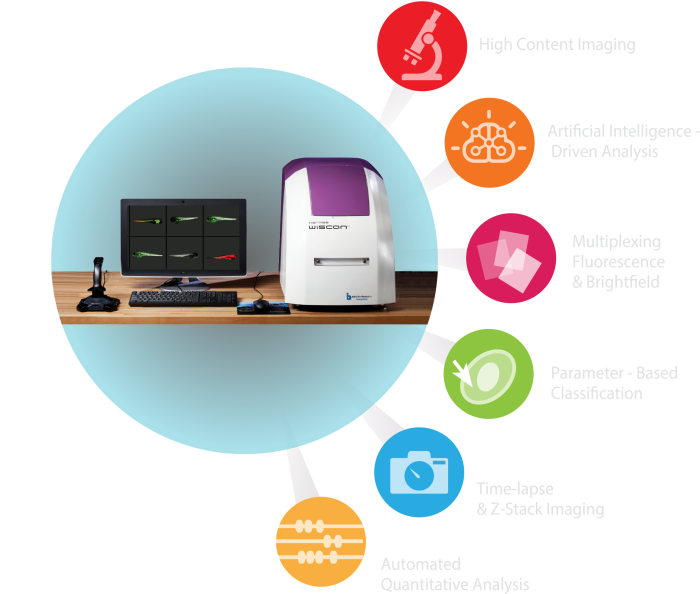
The Hermes automated high content imager from Idea-Bio is designed for analysis of 3D cultures (organoids and spheroids), primary, fixed and living cells. The Hermes is also well known for its easy and precise detection of zebrafish.

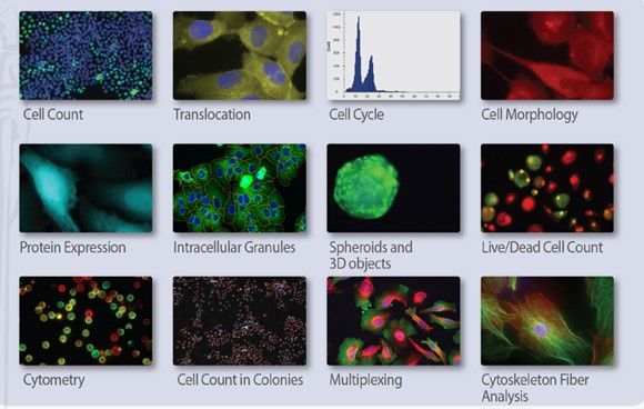
The system documents the quantification of your cells and its morphological changes. Also, it measures the fluorescence intensity.

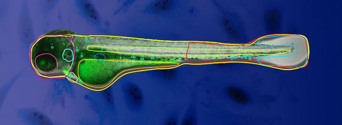
An specific example can explained with the zebrafish. The imaging of zebra fish is possible because of the automatically distinguishment of the parts of the fish (head, torso, tail) and its organs (including eye, yolk, spine, tail, brain, internal granules and more). With this feature the detection is eased as only the specific area of interest is analysed. This is all possible due to the Hermes artificial intelligence (AI) driven analysis.

HERMES benefits and features

Patented well scanning and smart software - Save time and eliminate errors

The Hermes High Content Imager has a patented U-shaped well scanning. This U-shaped well will place your cells and fish in the correct position.

Save time: The software automatically removes the images where the cells or organisms are not placed properly. 

  • Capture of >10 images per second
  • It is compatible for high throughput research.
  • Analyse multi well plates up to 1536 wells
  • Use slides, dishes of 35 mm and microarrays  
Hermes

Do you research with living cells? The HERMES environmental control makes it ideal to work with living cells.

  • Temperature can be set up to 40 °C
  • CO2 level can be set from 0.02 L/min to 0.13 L/min and adjusted from 0% to 15% CO2
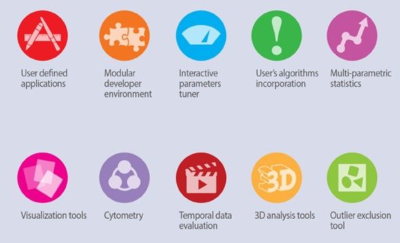
  • Customize the software to your research.


Optional Taylor-made software development

for specialized biological applications upon purchase

Contact us

zebrafish microscopy

LEDs and filters - HERMES captures brightfield and fluorescence images

HERMES presents 7 fluorescent colours and many air objectives. These 7 LED sources are DAPI, CFP, GFP, YFP, RFP, mCherry and CY5 plus a white LED. Besides that, it contains 2 emission filters and a compatible dichroic filter. The images that are retrieved are publicity quality, due to the varied zoom from 2x to 60x for the most detailed pictures.

Turn on Hermes imager  24/7

The fully automated high content imager requires minimal involvement. It loads and unloads the plates automatically into the imager and from the built-in stacker (20 plates). These plates can be incubated at 37 °C and 5% CO2. Additionally, it produces multiple visualisation tools such as scatter plots, histograms, heat maps, time graphs and dose curves.


Benefits of the WiScan Hermes:

  • Intuitively operated – images retrieved just by a push of a button
  • High speed acquisition - >10 images per second
  • Variable zoom and XYZ plane analysis
  • Correct placement of the cells


Athena Zebrafish Image Analysis Software

Revolutionary AI-based image analysis software for Zebrafish 

We present the Athena Zebrafish – the next generation in Zebrafish assays. Our new, unique dedicated analysis software for automated analysis of Zebrafish microscopy images offers simple and quick quantification of fluorescence, measurement of morphological changes & other phenotypic features in Zebrafish larvae in a high throughput format.

The software permits parameter-free zebrafish analysis using simple bright-field images. It automatically detects zebrafish embryos and larvae up to 5 days old (dpf), extracting the fish contour and much of its internal anatomy: yolk sac, eye, notocord, and more, along with body regions of the head, trunk, and tail. You can install it via this link!

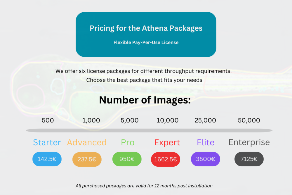
For analyzing images of proprietary formats (*.czi, *.nd2, etc) a one-time for lifetime license registration fee of $260.00 is required.

Order now!

Applications of the high content imager:


WiSoft

  • (Bacterial) Cell counting
  • Gene therapy
  • Organoid imaging
  • Phagocytosis
  • Cell adherence
  • Characterising disease mechanisms
  • Protein aggregation / accumulation assay
  • Stem cells imaging

For instance, studies have been performed using colon cancer stem cells, Dental Pulp stem cells  and Adipogenically Differentiating Mesenchymal Stromal/Stem Cells.

As research goes on, many more applications are supported by Hermes.

Do you want to know more information about it? 

CONTACT US

Request a quotation

Request a quotation for your selection of laboratory instruments, reagents, kits or consumables. We will get back to you via mail or phone in no more than 24 hours. Promised!

Do you want to know what we do with your data from this form? Then read our privacy statement.

Contact information

+31 30 6880771
support@isogen-lifescience.com

Our office

Veldzigt 2A
3454 PW De Meern

ISOgen office