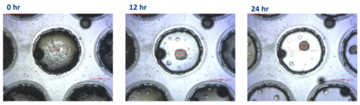
Live cell imaging system

Cell Imaging Systems

From routine cell monitoring to automated live cell analysis

Choosing the right cell imaging system depends on your research question. Do you need to quickly check cell morphology and confluency, or do you want to follow living cells continuously over time?

We offer cell imaging solutions for both routine cell culture observation and advanced live cell analysis. Whether you are documenting cell health, monitoring proliferation, studying migration, analysing immune cell killing, or following spheroid and organoid development, we help you find the imaging system that fits your workflow.

Explore our live cell imaging solutions:
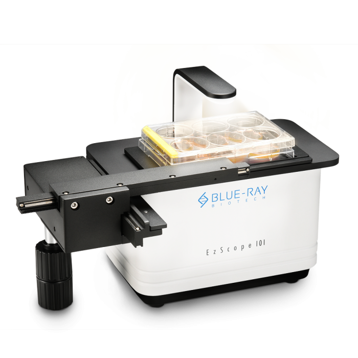
EzScope: Microscope-in-Incubator

Routine live cell monitoring without removing your cells from the incubator

Ideal for researchers who want to observe and document living cells while maintaining stable culture conditions. It allows you to monitor morphology, confluency, and cell health directly from inside the incubator, reducing manual handling and helping you follow your cultures more easily.

Keep your cells alive through the entire process of your research!

Cell culture enables you to grow cells for a specific period to study cells behaviour under specific conditions depending on each researcher's life science project. However, what all of them have in common is checking cell's growth health. Cells need to grow happy and healthy. 

 With our new microscope-in-incubator solution, the hassles of removing cells from the incubator for observation disappear. Incorporate the microscope inside the incubator and visualise your cells from your computer. Therefore, you can see if your cells need new media or not, how they are growing, and even visualise cell migration or aggregation without even standing up!

Capture your cells in real-time

Being able to monitor your cells without the need of taking them out of the incubator enables to perform studies such as cell migration, growth, and invasion between other phenotypic cell-based assays.

Live cell imaging systems help to streamline your research workflow with improved efficiency and productivity. EzScope 101 brings 24/7 measurements under precisely controlled conditions in a non-perturbing environment. You can observe the images anytime, with walk-away convenience.  Up to four samples can be monitoring simultaneously in a same incubator. This feature helps reduce repetitive action, saves time, and op timizes experiment efficiency.

The EzScope in your lab benefits your research!

  • Error reduction: Less errors because of a decrease in environmental variations due to a non-perturbing environment.
  • Space saving: Its compact design in size enables you to connect up to 4 units.
  • Reduce contamination risk
  • Save time and optimize experiment efficiency thanks to monitoring up to four samples in the same incubator.
  • EzCapture Software enables easy images editing, correction of background brightfield, and creation of time-lapse video output.
  • Your cells, from anywhere: Remote monitoring of your cell asssays.
  • Maintenance based on minimal cleaning 

Assay Applications

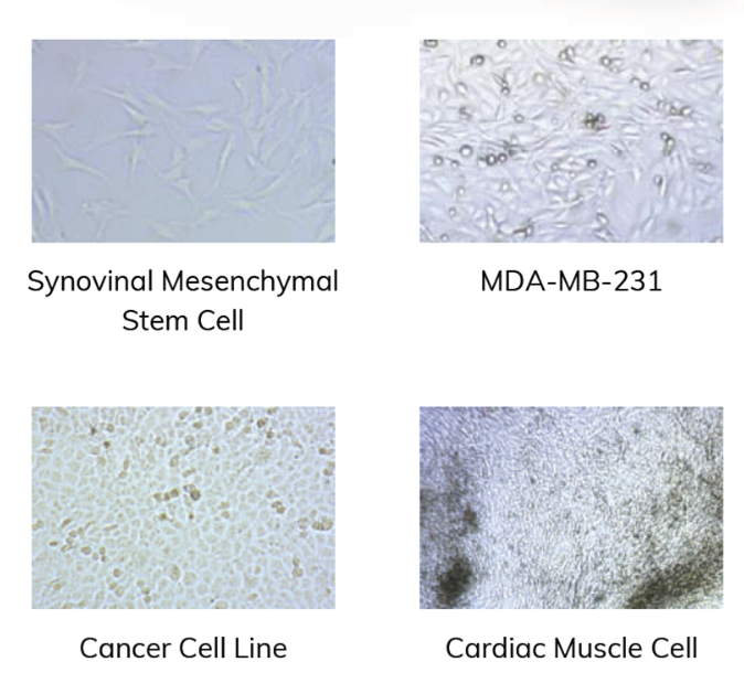
EzScope has recorded different cell lines for different projects worldwide successfully. The broad range of applications where it can be used is listed below.

  • Catch me if you can! Recognition and attachment of human killer T cells to cancer cells to kill them, Cancer treatment drug screening, Immunotherapy research, Chemotaxis 
  • Cell migration: Tumor Cell Cluster formation, wound healing, cell confluence... 
  • Stem Cell Research: monitor cell behavior when inoculation on a gelatin matrix. 
  • Pathology infection experiments: For instance, the determination of infection can be performed by analysing morphology changes.
  • NEW! Discovering genes and microRNAs involved in human lung development unveils IGFBP3/miR-34a dynamics and their relevance for alveolar differentiation

User reviews

Customer review

Differentiation of human embryonic Lung Organoids

The EzScope has been a valuable tool in our laboratory. This imaging microscope for incubators has allowed us to accurately capture the differentiation process of human embryonic lung organoids into alveolospheres-like structures following transfection with IGFBP3 siRNA.

Documenting morphological changes at regular intervals without disrupting the incubator environment has been crucial for our analysis. The ease of use and the quality of the images obtained with the EzScope have improved the productivity of our experiments, enabling continuous and precise monitoring of organoid development.

Laboratory of Molecular Oncology and Embryology

Spica M1: Automated In-Incubator Live Cell Imaging

Continuous live cell imaging with automated quantitative analysis

Spica M1 is designed for researchers who need more than visual observation. It enables automated image acquisition inside the incubator, combining brightfield and fluorescence imaging with AI-assisted analysis to generate quantitative data over time.

Use it to monitor dynamic biological processes such as proliferation, migration, cytotoxicity, transfection efficiency, spheroid growth and organoid development.


Benefits

Keep your cells where they belong

Image living cells directly inside the incubator without disturbing temperature, humidity or CO₂ conditions. By minimizing manual handling, experiments remain more reproducible while preserving cell health throughout long-term studies.

Follow dynamic biology in real time

Capture the complete story behind your experiment instead of a single endpoint. Automatically monitor cell proliferation, migration, immune cell interactions, transfection efficiency and 3D culture development over hours or days, generating continuous quantitative data as your experiment progresses.

Brightfield and fluorescence in one platform

Visualize living cells using label-free brightfield imaging or combine up to three fluorescence channels to study multiple biological processes simultaneously. From simple morphology assessments to complex multi-marker experiments, Spica M1 adapts to your research needs.

AI-assisted image analysis

Move beyond image acquisition. Integrated AI-powered analysis automatically transforms images into meaningful biological data, allowing you to quantify proliferation, wound healing, cytotoxicity, transfection efficiency and spheroid growth with minimal manual intervention. Custom AI training also enables accurate analysis of challenging samples.

Unparalleled Flexibility for Diverse Live Cell Assays


Applications

The Spica M1 supports a broad range of live cell applications, including:

  • Cell proliferation and growth kinetics
  • Immune cell killing assays (CAR-T, NK cells and co-cultures)
  • Wound healing and migration assays
  • 3D spheroid and organoid analysis
  • Transfection efficiency
  • Long-term live cell monitoring
  • Label-free and fluorescence imaging

Ready to transform your live cell imaging workflow?

Whether you are following organoid development, evaluating immunotherapy responses or monitoring long-term cell behaviour, the Spica M1 delivers automated imaging and quantitative analysis without disrupting your experiment.

✔ Request a live demonstration

Request a quotation

Request a quotation for your selection of laboratory instruments, reagents, kits or consumables. We will get back to you via mail or phone in no more than 24 hours. Promised!

Do you want to know what we do with your data from this form? Then read our privacy statement.

Contact information

+31 30 6880771
support@isogen-lifescience.com

Our office

Veldzigt 2A
3454 PW De Meern

ISOgen office